.
Thereof, how does the orbicularis oculi work?
The orbicularis oculi muscle lies directly underneath the surface the skin, around the eyes. Its function is to close the eyelid, and to help in the passing and draining of tears through the punctum, canaliculi, and lacrimal sac, all parts of the tear drainage system.
Also Know, how did the orbicularis oculi get its name? The English name for this muscle is the little circle muscle of the eye. The word orbicularis comes from the Latin orbis meaning “circle or disk” and ulus which is a diminutive ending meaning “little.” The word oculi is from the Latin oculus meaning “eye.”
Then, what type of muscle is the orbicularis oculi?
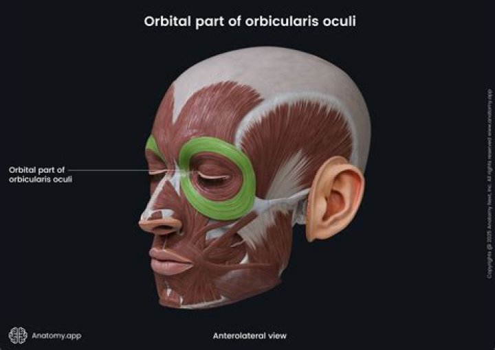
Shape. The orbicularis oculi muscle is a broad, flat, sheet of skeletal muscle with orbital, palpebral and lacrimal portions. The circular orientation of the fibres is a reflection of the sphincter-like function of this muscle.
Where is the orbicularis oculi muscle?
The orbicularis oculi is a muscle in the face that closes the eyelids. It arises from the nasal part of the frontal bone, from the frontal process of the maxilla in front of the lacrimal groove, and from the anterior surface and borders of a short fibrous band, the medial palpebral ligament.
Related Question AnswersHow do you test for orbicularis oculi?
Test the strength of the orbicularis oculi by gently trying to pry open the patient's upper eyelid. Instruct him to puff out both cheeks. Check tension by tapping his cheeks with your fingers. Have the patient smile broadly and show his teeth, testing the lower face.What is the kissing muscle called?
Located in the face, the orbicularis oris muscle controls movements of the mouth and lips. The muscle inserts directly into the lips. In common language, the orbicularis oris is often referred to as 'the kissing muscle.What is the smiling muscle?
The zygomaticus major is a muscle of the human body. It is a muscle of facial expression which draws the angle of the mouth superiorly and posteriorly to allow one to smile.What muscle opens and closes the eye?
orbicularis oculiHow many muscles does it take to open your eyes?
6 musclesWhat is the inside of your eyelid called?
Conjunctiva The conjunctiva makes up the lining inside your eyelids. It almost entirely covers your sclera, and is nourished by tiny blood vessels that are almost invisible to the naked eye.What nerve opens the eye?
oculomotor nerveWhere is the Buccinator muscle located?
The buccinator (/ˈb?ks?ne?t?r/) is a thin quadrilateral muscle occupying the interval between the maxilla and the mandible at the side of the face. It forms the anterior part of the cheek or the lateral wall of the oral cavity.Where is the Platysma muscle located?
Platysma Muscle. The platysma muscle is a broad sheet of muscle arising from the pectoral (chest) and deltoid (shoulder) muscles and rises over the collarbone (clavicle), proceeding upward in a slanting manner along the sides of the neck.Where is the Zygomaticus muscle located?
Zygomaticus major. The zygomaticus major muscle is a muscle that controls facial expression, drawing the mouth's angle upward and outward. The zygomaticus major muscle starts at the cheekbone and extends to the corner of the mouth. This muscle causes the corners of a person's mouth to rise when they smile.What muscle closes the jaw?
The masseter elevates the jaw, closing the mouth. The temporalis elevates and retracts the jaw. The lateral pterygoid is the only muscle of mastication that actively opens the jaw.How do your eyelids work?
An eyelid is a thin fold of skin that covers and protects an eye. The levator palpebrae superioris muscle retracts the eyelid, exposing the cornea to the outside, giving vision. They keep the eyes from drying out when asleep. Moreover, the blink reflex protects the eye from foreign bodies.What happens if you damage your temporalis?
The temporalis muscle can sustain injury leading to atrophy by denervation of the muscle, damage to its vascular supply, or direct muscle fiber injury.What muscles move your eyelids?
The lids move through the action of a circular lid-closing muscle, the orbicularis oculi, and of the lid-raising muscle, the levator of the upper lid.What is the temporalis muscle?
The temporal muscle, or temporalis muscle, is one of several chewing muscles that is necessary for crushing and grinding objects between the molars. Due to its location and frequent use, this muscle may be a primary focal point for a recurring condition known as “tension headaches.”What muscle allows you to frown?
Orbicularis oculiWhat muscles are named for their size?
Size: Maximus (largest), minimus (smallest), longus (longest), and brevis (shortest) are common suffixes added to muscle names.What are the 7 ways muscles are named?
- Location. ex. Rectus Abdominis.
- Action. ex. Extensor Digitorum Longus.
- Direction of Fibers. ex. Extrenal Oblique.
- Shape. ex. Deltoid.
- Number of Origins. ex. Triceps Brochii.
- Origin/Insertion. ex. Sternocleidomastoid.
- Relative Size. ex. Adductor Magnus.